39 diagram of sheep brain
Androgens increase excitatory neurogenic potential in ... 19-01-2022 · The biological basis of male–female brain differences has been difficult to elucidate in humans. The most notable morphological difference is size, with male individuals having on average a ... DOC Sheep Brain Anatomy Lab Manual - amherst.edu Sheep Brain Anatomy Lab Manual. Based on original material by R. N. Leaton, Dartmouth College. Contributors to this version: Al Sorenson, Lisa Raskin, Sarah Turgeon, Steve George, and JP Baird. I. Introduction. The brain of the sheep is useful for study because its anatomy is similar to human brain anatomy. Although exact proportions (and names ...
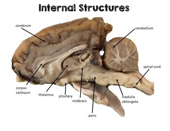
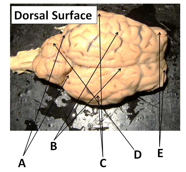
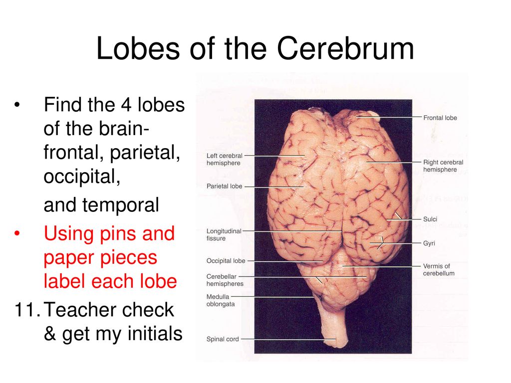
Sheep Brain Dissection Lab The lobes of the brain are visible, as well as the transverse fissure, which separates the cerebrum from the cerebellum. The convolutions of the brain are also visible as bumps (gyri) and grooves (sulci). Use the diagram below to help you locate these items. Dorsal View of the Sheep Brain . 8.
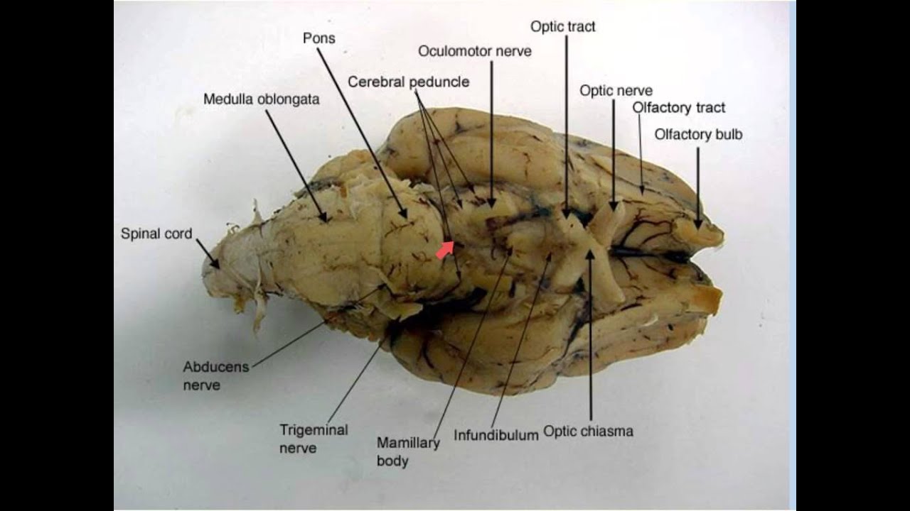
Diagram of sheep brain
Sheep Brain Dissection Project Guide | HST Learning Center Place the brain with the curved top side of the cerebrum facing up. Use a scalpel (or sharp, thin knife) to slice through the brain along the center line, starting at the cerebrum and going down through the cerebellum, spinal cord, medulla, and pons. Separate the two halves of the brain and lay them with the inside facing up. 2. Sheep Brain Anatomy Quiz - ProProfs Quiz Sheep Brain Anatomy Quiz. Sheep are wonderful and cute. The brain is an interesting organ. It helps with cognition and memory. Almost all the basic task In the body is commanded by the Brain. It is the control center of the body which regulates and control the process crucial for survival Are you interested in learning more about the brain of ... sheep-brain - Bay Path University 1. lateral ventricle: 11. fourth venticle: 2. fornix: 12. aqueduct: 3. corpus callosum: 13. pons: 4. pineal body: 14. pituitary gland: 5. superior colliculi: 15 ...
Diagram of sheep brain. Sheep Brain Map: Midsaggital view This map shows the major structures of the sheep brain with an active cursor to help identify the structures Neuroanatomy Tutorial I: Basic Anatomy of the Brain Point to any region of this midsaggital sheep brain image (medial view) to highlight that structure. Click the left mouse button to identify the structure you are pointing to. Elephant Anatomy Facts - Complete Diagram Of Anatomy The brain weight of the male African elephant is 4.2-5.4 kilograms. The brain weight of the female African elephant is 3.6 – 4.3 kilograms. Both are quite heavy in comparison to the adult human brain although brain development in elephants is quite similar to that of human beings. Humans are born with small brain mass, so are elephants. sheep brain Diagram | Quizlet sheep brain Diagram | Quizlet sheep brain STUDY Learn Write Test PLAY Match Created by haley_head6 Terms in this set (23) arbor vitae ... inferior colliculus ... pons ... spinal cord ... cerebellum ... 4th ventricle ... thalamus ... hypothalamus ... cerebral aqueduct ... habenula ... medulla oblongata ... optic chiasma ... tela choroidea ... fornix Brain Diagram Pituitary Gland - sheep brain dissection lab ... Brain Diagram Pituitary Gland - 14 images - the human brain its parts and functions, sheep brain dissection bi biology junction, endocrine glands openstax biology, 2013 types of tissue and glands composing endocrine system,
Medulla oblongata - Wikipedia The base of the medulla is defined by the commissural fibers, crossing over from the ipsilateral side in the spinal cord to the contralateral side in the brain stem; below this is the spinal cord. Blood supply. Blood to the medulla is supplied by a number of arteries. Sheep Brain Anatomy and Function Flashcards - Cram.com An extremely efficient memory center in the brain. Responsible for spatial memory and spatial navigation as well as some types of non-spatial memory such as contextual memory and episodic memory. Damage to this structure produces both anterograde and retrograde amnesia. - Vertical cut through pineal gland laterla to pineal gland. PDF Lab: Sheep Brain Dissection - Mrs. Moretz's Science Site Lab: Sheep Brain Dissection 1 **Before starting this lab, open the "Brain Parts and Functions" document. Refer to images, descriptions, and functions of parts of the brain as you proceed through this lab. Sheep brains, although much smaller than human brains, have similar features and can be a valuable addition to anatomy studies. Human vs Sheep Brain [classic] | Creately Human vs Sheep Brain [classic] Use Creately's easy online diagram editor to edit this diagram, collaborate with others and export results to multiple image formats. You can edit this template and create your own diagram. Creately diagrams can be exported and added to Word, PPT (powerpoint), Excel, Visio or any other document.
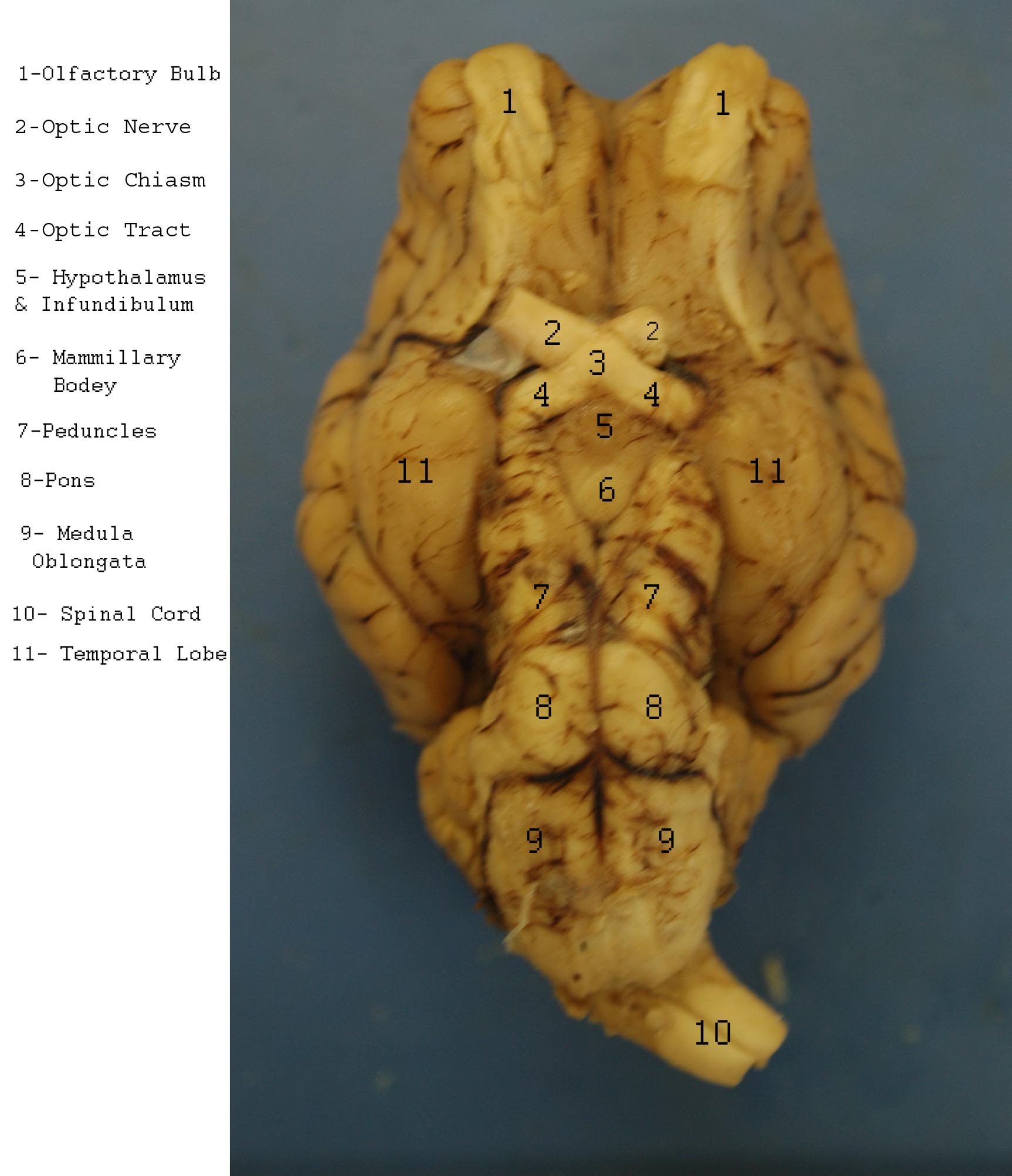
Diagram of Sheep Brain - Inferior view Diagram of Sheep Brain - Inferior view Technology at MSU - Andrew File System Retirement | Michigan ... Andrew File System (AFS) ended service on January 1, 2021. AFS was a file system and sharing platform that allowed users to access and distribute stored content. AFS was available at afs.msu.edu an… PDF Sheep Brain Dissection Lab - Home Science Tools Place the brain with the curved top side of the cerebrum facing up. Use a scalpel (or sharp, thin knife) to slice through the brain along the center line, starting at the cerebrum and going down through the cerebellum, spinal cord, medulla, and pons. Separate the two halves of the brain and lay them with the inside facing up. 2. Sheep Brain & Eye (with labels) - YouTube A video tutorial of the anatomy of the brain and eye of a sheep for comparative anatomy. A video tutorial of the anatomy of the brain and eye of a sheep for comparative anatomy.
sheep brain anatomy
Recombinant DNA Technology (With Diagram) Biotechnologists have successfully produced transgenic pigs, sheep, rats and cattle. (3) Production of Hormones: By the advent of techniques of rec DNA technology, bacterial cells like E.coli are utilized for the production of different fine chemicals like insulin, somatostatin, somatotropin and p-endorphin.

Resources for Teaching Mammalian Neuroanatomy Using Sheep ...
Diagram of Sheep Brain - Lateral view - Modesto Junior College Diagram of Sheep Brain - Lateral view - Modesto Junior College
Index of /files/OCC_VIDEO/upload/Faculty_Resources/acamilo ...
Sheep Brain - San Diego Mesa College Sheep Brain. Click on a photo for a larger view of the model. Click on Label for the labeled model. Back to Dissected Specimen Page. Superior View. Corpora Quadrigemina. Label.
SCB209 - Lab2 - Natural Sciences Open Educational Resources
11.7: Sheep Brain Dissection - Biology LibreTexts The sheep brain is enclosed in a tough outer covering called the dura mater. You can still see some structures on the brain before you remove the dura mater. Take special note of the pituitary gland and the optic chiasma. These two structures will likely be pulled off when you remove the dura mater. Figure 11.7. 1: Brain with Dura Mater Intact
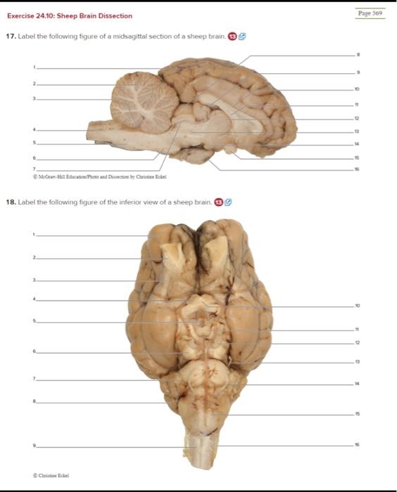
Solved Exercise 24.10: Sheep Brain Dissection Page 509 ...
Sheep Brain Dissection | Carolina.com Carolina's Perfect Solution® sheep brain dissection introduces students to the anatomy of a mammalian brain, both external and internal, and encourages students to construct an explanation of the central nervous system. Below is a brief survey of the internal and external anatomy of the sheep brain.

Diagram of Sheep Brain - Inferior view
CDC - Echinococcosis - Biology However, genotypes G1 and G3 (associated with sheep) are the most commonly reported at present and broadly distributed. In North America, Echinococcus granulosus is rarely reported in Canada and Alaska, and a few human cases have also been reported in Arizona and New Mexico in sheep-raising areas. In the United States, most infections are ...
Nervous System Activity - Sheep Brain Dissection
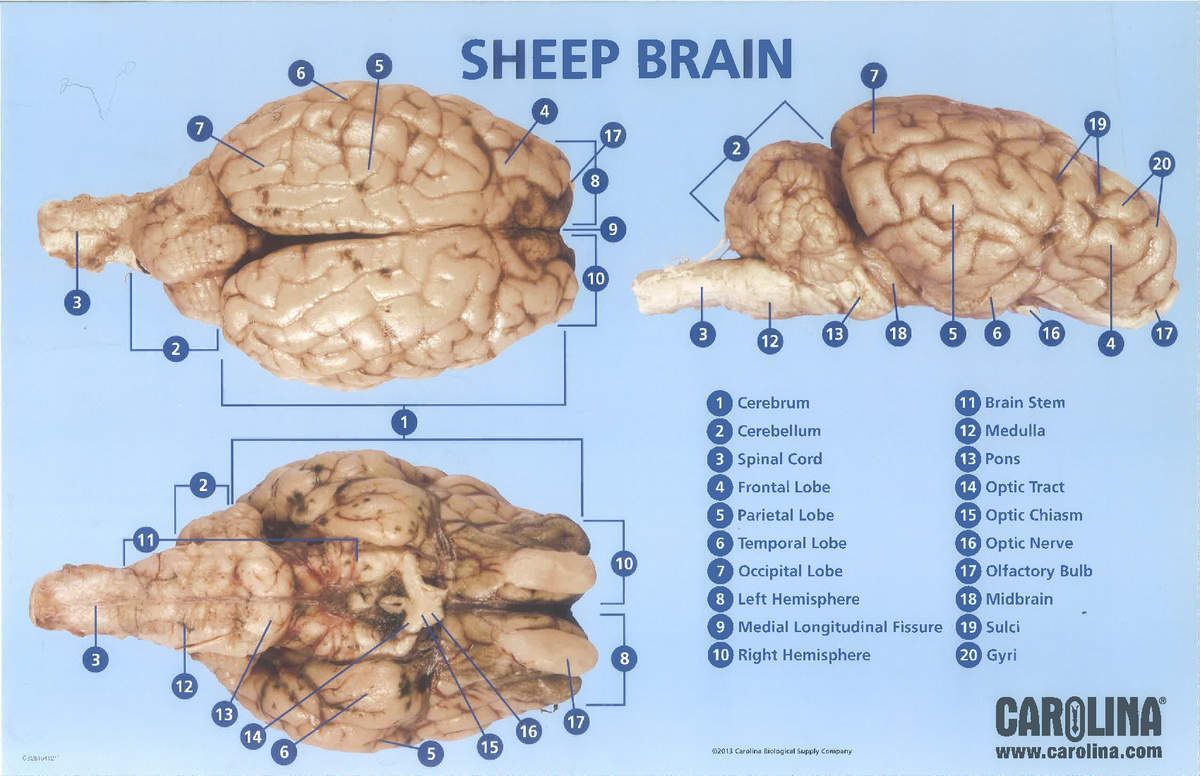
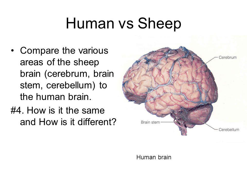
Difference Between Human and Sheep Brain (With Table ... A sheep 's brain weighs the tenth part of the human brain. The human brain is 15 centimetres long and a sheep 's brain is only about a third of that length. Both have three divisions namely, cerebrum, cerebellum and the brainstem. The cerebellum and brain stem are located behind the cerebrum in sheep as they have a horizontal spine.
Sheep Brain Dissection Report
Sheep Brain Dissection Guide with Pictures | Nervous ... Sheep Brain Dissection Guide. Dissection guide with instructions for dissecting a sheep brain. Checkboxes are used to keep track of progress and each structure that can be found is described with its location in relation to other structures. An image of the brain is included to help students find the structures. spaceandmystery.
DISSECTION OF THE SHEEP'S BRAIN
Solved Art-labeling Activity: Midsagittal Section of the ... Anatomy and Physiology questions and answers. Art-labeling Activity: Midsagittal Section of the Sheep Brain (Diagram, 2 of 2) Reset Help Fomix Infundibulum Olfactory bulb Optic chiasm Mosencephalon Pituitary gland Marillary body Medulla oblongata Pons Spinal cord Corpus callosum Art-labeling Activity: Midsagittal Section of the Sheep Brain ...
Sheep Brain Neuroanatomy Online Self-Test | KPU.ca - Kwantlen ...
Prefrontal cortex - Wikipedia In mammalian brain anatomy, the prefrontal cortex (PFC) is the cerebral cortex which covers the front part of the frontal lobe.The PFC contains the Brodmann areas BA8, BA9, BA10, BA11, BA12, BA13, BA14, BA24, BA25, BA32, BA44, BA45, BA46, and BA47.. The basic activity of this brain region is considered to be orchestration of thoughts and actions in accordance with …
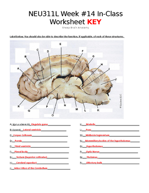
Week 14 worksheet Sheep brain neuroanatomy Online S20 KEY ...
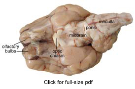
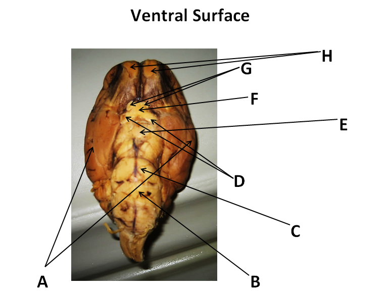
Sheep brain dissection | Human Anatomy and Physiology Lab ... The most prominent structure visible on the ventral side of the sheep brain is half of the optic chiasma, which is where the two optic nerves cross over each other and form an "X" shape. You will only see half the structure. Find the optic chiasma half on your brain. You may have removed the optic removed the chiasma with the dura mater.

One hemisphere of sheep brain that is cut into four blocks ...
Sheep Brain - Dorsal View Brain Anatomy IntroductionCLOSE Dissected Sheep Brain — Dorsal View Dorsal view of sheep brain with the cerebellum and caudal cerebrum removed. The rostral colliculus(large arrow label) and the caudal colliculus(small arrow label) together form the tectumof the midbrain.

Sheep Brain Sagittal View Diagram | Quizlet
Sheep Brain Dissection with Labeled Images Sheep Brain Dissection 1. The sheep brain is enclosed in a tough outer covering called the dura mater. You can still see some structures on the brain before you remove the dura mater. Take special note of the pituitary gland and the optic chiasma. These two structures will likely be pulled off when you remove the dura mater.

Sheep brain exterior | Brain science, Life science middle ...
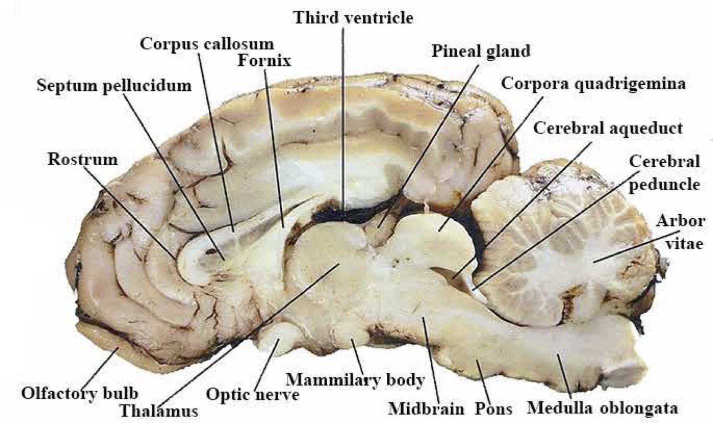
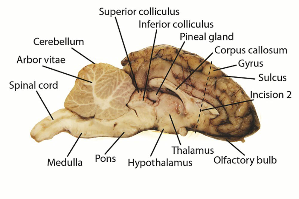
PDF Sheep Brain Midsagittal Section - Dr. Scott Croes' Website Sheep Brain -Parasagittal Section 1. Gray Matter 2. White Matter 3. Corpus Callosum 4. Lateral Ventricle 5. Caudate Nucleus 6. Septum Pellucidum 7. Fornix 8. Optic Chiasma 9. Third Ventricle 10. Thalamus (Ovid Nuclear Mass of Thalamus) 11. Corona Radiata 12. Hippocampus 13. Cerebral Aqueduct (of Sylvius) 14. Pituitary Gland (hypophysis) 15.

Drawing of a midsagittal section of the sheep brain ...
Sheep Brain Neuroanatomy Online Self-Test | KPU.ca ... Sheep Brain Neuroanatomy Online Self-Test. Use each diagram as a reference, and selected the correct answer for each lettered structure. You may find it useful to open the diagrams in a separate window to review while answering each question.

inferior view of sheep brain
PDF DISSECTION OF THE SHEEP'S BRAIN - Hanover College Sheep Brain Dissection Guide Planes of Orientation In addition to the direction, the brain as a three dimensional object can be divided into three planes. There is the frontal or coronal planes which divides front from back. It can divide the brain and any location as long as it divides the brain from front to back.
Sheep Brain Dissection Guide - ppt download
Circular RNA - Wikipedia Circular RNA (or circRNA) is a type of single-stranded RNA which, unlike linear RNA, forms a covalently closed continuous loop. In circular RNA, the 3' and 5' ends normally present in an RNA molecule have been joined together. This feature confers numerous properties to circular RNA, many of which have only recently been identified. Many types of circular RNA arise from …

Sheep Brain V (Lateral view) Diagram | Quizlet
SHEEP BRAIN IMAGES - sdmesa.edu NERVOUS SYSTEM - SHEEP BRAIN IMAGES. Sheep Brain Unlabeled. Sheep Brain Leader-Lined. Sheep Brain Labeled. San Diego Mesa College 7250 Mesa College Drive San Diego, CA 92111-4998 Student Support San Diego Community College District San Diego City College San Diego Mesa College San Diego Miramar College San Diego Continuing Education.

Adventures with animals and plants. Biology. Fig. 255 A ...
Sheep Heart Dissection Lab for High School Science | HST Most heart diagrams show the left atrium and ventricle on the right side of the diagram. Imagine the heart in the body of a person facing you. The left side of their heart is on their left, but since you are facing them, it is on your right. 1. Identify the right and left sides of the heart.
Lab: Sheep Brain Dissection
Prions - PMC Prion protein isoforms. (A) Western immunoblot of brain homogenates from uninfected (lanes 1 and 2) and prion-infected (lanes 3 and 4) Syrian hamsters.Samples in lanes 2 and 4 were digested with 50 µg/µl proteinase K for 30 min at 37°C, completely hydrolyzing PrP C.Proteinase digestion cleaves ∼67 amino acids from the amino terminus of PrP Sc to generate PrP 27–30 …

File:Sheep Brain Dissection 2 - black background.png - Wikipedia
PDF Neuroanatomy: Dissection of the Sheep Brain Examine the sheep brain with the membranes intact. You should be able to identify and use the following directional terms: Anterior / Posteriorfront / back Rostral / Caudal towards the beak / towards the tail Medial / Lateral towards the middle / towards the side Dorsal / Ventral top / bottom (on the CNS of a quadruped)
Sheep Brain Dissection Project Guide | HST Learning Center
What is Menstrual Cycle: Phases, Diagram, Reasons - Embibe Apr 03, 2022 · The menstrual cycle is the term related to cyclic changes that occur in the reproductive system of the primate female’s body every month, while oestrous cycle is the term used to describe cyclic changes that occur in the reproductive system of the non-primate (cow, sheep, buffalo, etc.) female’s body every month. Learn Exam Concepts on Embibe

Cerebrum Sheep Dissection - Human Anatomy - GUWS Medical
sheep-brain - Bay Path University 1. lateral ventricle: 11. fourth venticle: 2. fornix: 12. aqueduct: 3. corpus callosum: 13. pons: 4. pineal body: 14. pituitary gland: 5. superior colliculi: 15 ...

ImageQuiz: Sheep brain dorsal view
Sheep Brain Anatomy Quiz - ProProfs Quiz Sheep Brain Anatomy Quiz. Sheep are wonderful and cute. The brain is an interesting organ. It helps with cognition and memory. Almost all the basic task In the body is commanded by the Brain. It is the control center of the body which regulates and control the process crucial for survival Are you interested in learning more about the brain of ...
Sheep brain dissection - Bisc 163 - StuDocu
Sheep Brain Dissection Project Guide | HST Learning Center Place the brain with the curved top side of the cerebrum facing up. Use a scalpel (or sharp, thin knife) to slice through the brain along the center line, starting at the cerebrum and going down through the cerebellum, spinal cord, medulla, and pons. Separate the two halves of the brain and lay them with the inside facing up. 2.

Sheep Brain Dissection Lab Companion

Sheep Brain Dissection | Carolina.com
Sheep Brain Neuroanatomy Online Self-Test | KPU.ca - Kwantlen ...

Sheep Brain Images
Sheep Brain Dissection Project Guide | HST Learning Center

Sheep brain images | Lab | Amherst College
Sheep Brain Dissection
Sheep Brain Dissection Guide - ppt video online download
Topic: Sheep Brain Dissection Grades: 8-12th Number of ...

HSF Lab 1 - Exercise 17 (sheep brain) Diagram | Quizlet

Sheep Brain Images
Medical Detectives Lesson 27
Sheep Brain Dissection

Sheep Brain Images
Comments
Post a Comment