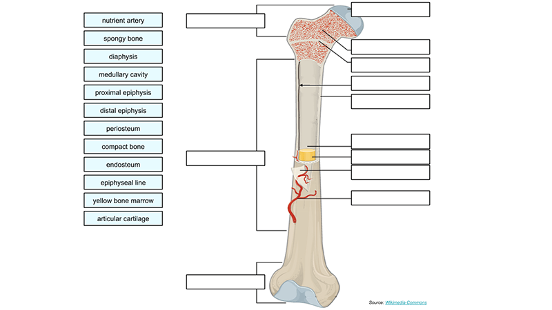
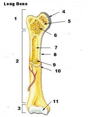
43 long bone diagram unlabeled
Long bones are one of the five bone types that are classified by shape. Long bones include the humerus (upper arm), radius (forearm), ulna (forearm), femur (thigh), fibula (thin bone of the lower leg), tibia (shin bone), phalanges (digital bones in the hands and feet), metacarpals (long bones within the hand), and metatarsals (long bones within the feet). Besides having a significant length vs width when compared to most other bones, long bones are also responsible for supporting weight and ... its unlabeled, so that your practce better. carotid canal coronal suture ethmoid bone external occipital protuberance foramen lacerum foramen magnum foramen
2XL/3XL. $62.00. Quick Shop. BONE SOCKS WITH TAUPE LOGO. S/M. M/L. $12.00. Elevated staples meet lux fabrications for a versatile and comfortable wardrobe that simplifies and amplifies your day to day wear. When you purchase something from Unlabeled it has a tag that says "Remove label before wearing" This message was purposefully placed as a ...

Long bone diagram unlabeled
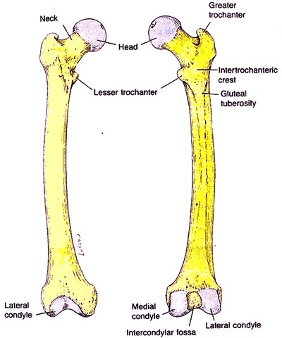
Unlabeled diagram showing the femur (download free PDF below!) ... Basic anatomy and parts of the femur. The femur is a long bone found in the lower extremity. It serves as the attachment site for several muscles of the hip and leg, allowing it to withhold pressure from multiple angles. There are three main parts to the femur: The proximal end; Humerus bone labeled vector illustration diagram stock illustration. Save to Board. Humerus bone labeled vector illustration diagram Humerus bone labeled vector illustration diagram. Long bone type in the upper arm. Skeleton anatomy scheme with greater tubercle, deltoid tuberosity, medial epicondyle, trochlea and other parts. Humerus stock vector. Femur Bone Anatomy. The femur is a type of long bone located in the thigh and is the largest bone of the skeletal system. The femur and/or hip may fracture secondary to trauma, so understanding the femur bone anatomy is important. The anatomy of the femur can be divided into proximal, central, distal, and posterior parts.
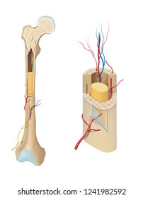
Long bone diagram unlabeled. In long bones, as you move from the outer cortical compact bone to the inner medullary cavity, the bone transitions to spongy bone. Figure 6.3.6 – Diagram of Compact Bone: (a) This cross-sectional view of compact bone shows several osteons, the basic structural unit of compact bone. (b) In this micrograph of the osteon, you can see the ... Diagram unlabeled skull diagram inferior view template information title unlabeled skull diagram inferior view categories diagram publised tuesday january 17th 2017 04 55 09 am. Long blank long bone diagram bone structure diagram and metaphysisjpg from the above resolutions which is part of the human anatomydownload this image for free in hd ... A quality educational site offering 5000+ FREE printable theme units, word puzzles, writing forms, book report forms,math, ideas, lessons and much more. Great for new teachers, student teachers , homeschooling and teachers who like creative ways to teach. Join the popular membership section!! Long Bone Diagram Unlabeled via. Tibia and Fibula Diagram Unlabeled via. Unlabeled Vertebral Column Diagram via. Cervical Vertebrae Blank Diagram via. Femur Bone Diagram Unlabeled via. Human Body Muscles via. Human Body Muscles via. This website is consists of people that are very appreciate creativity from every one, with no exception. That is ...
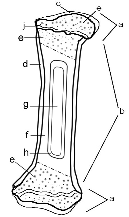
Image size: 25.0 Mpixels (71.6 MB uncompressed) - 5001x5001 pixels (16.6x16.6 in / 42.3x42.3 cm at 300 ppi) Published in: Bones, Joints and Muscles Images , Orthopaedics & Sport Medicine Images & Videos. Results for long bone diagram unlabeled. The humerus bone is a long bone that is part of the: Unlabeled skeleton print out 13 bones of the head and. Parts of a long bone unlabeled diagram system. Radius and ulna bone anatomy via. Each system contains haversian canals surrounded by concentric lamellae of bone tissue 48. 8 Jul 2015 — a = epiphysis b = diaphysis c = articular cartilage d = periosteum f = compact bone g = medullary cavity (yellow marrow) h = endosteum Start studying Unlabeled long bone. Learn vocabulary, terms, and more with flashcards, games, and other study tools.
Apr 14, 2019 · Long Bone Diagram Unlabeled ~ thanks for visiting our site, this is images about long bone diagram unlabeled posted by Alice Ferreira in Diagram category on Nov 02, You can also find other images like wiring diagram, parts diagram, replacement parts, electrical diagram, repair manuals, engine diagram, engine scheme, wiring harness, fuse box, vacuum diagram, timing belt, timing chain, brakes . Wedge-shaped segment from the compact part of a long bone 1. Periosteum 2. Outer general lamellae 3. Perforating fibers of Sharpey 4. Osteon with Haversian vessel Presentation of the spiral run of the collagenous fibrils within the single lamellae 5. Osteon w/presentation of the flattened bone cells 6. Bone cell 7. Branch of the bone cell 8. Oct 24, 2021 · Body Printable Blank Muscle Diagram. Labeled diagrams will be very important in a physiology classthe smartest organ in the body. Human Anatomy Rib Cage Muscles – 5 Best Images of Upper Limb Labeling Worksheet – Long Bone Diagram Unlabeled Human Body Muscles Jan 26 2013 human anatomy of muscles natural health newsletter. LABELING EXERCISE: BONES OF THE AXIAL AND APPENDICULAR SKELETON . Most, but not all, features you are required to know are shown on the following pages. Study from the bone list or your textbook after you marked the drawings as instructed on page 6-2. After you have studied the bones in lab, label the drawings as a self-test. Do not spend your
34 Label The Parts Of A Long Bone - Labels Design Ideas 2020
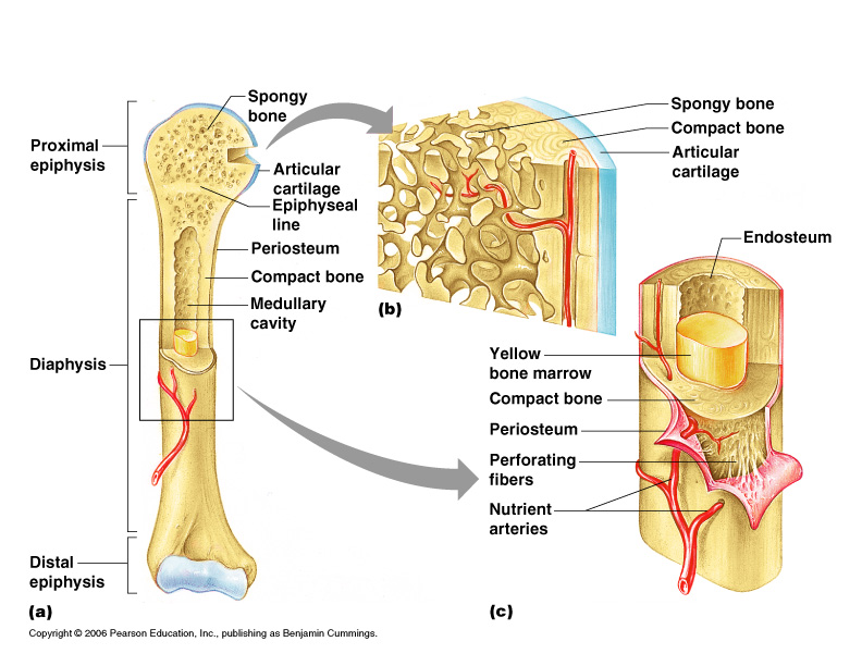
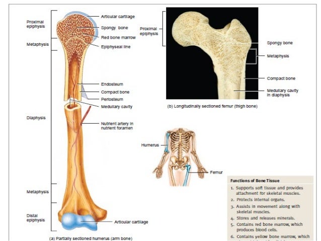
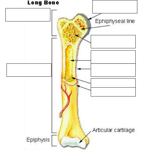
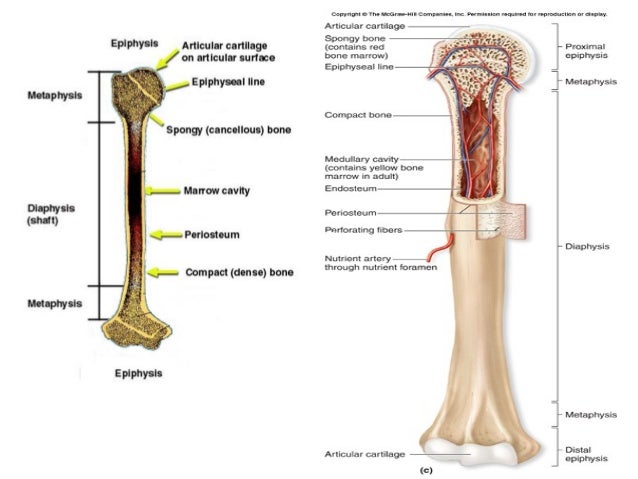
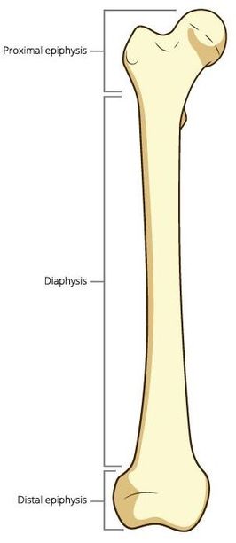
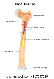
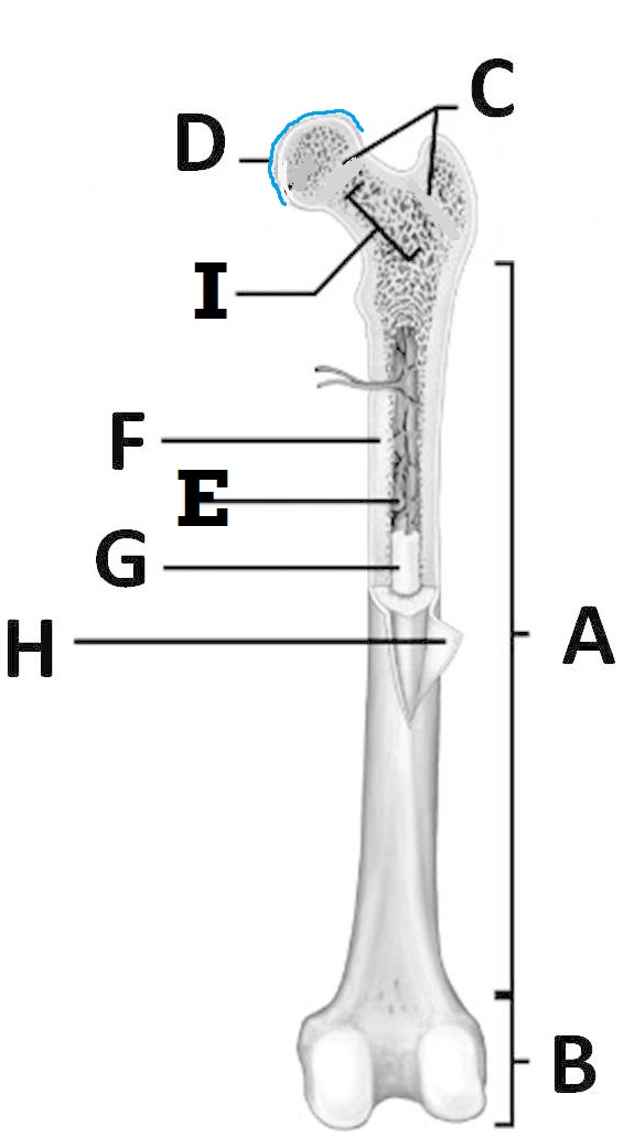
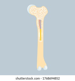
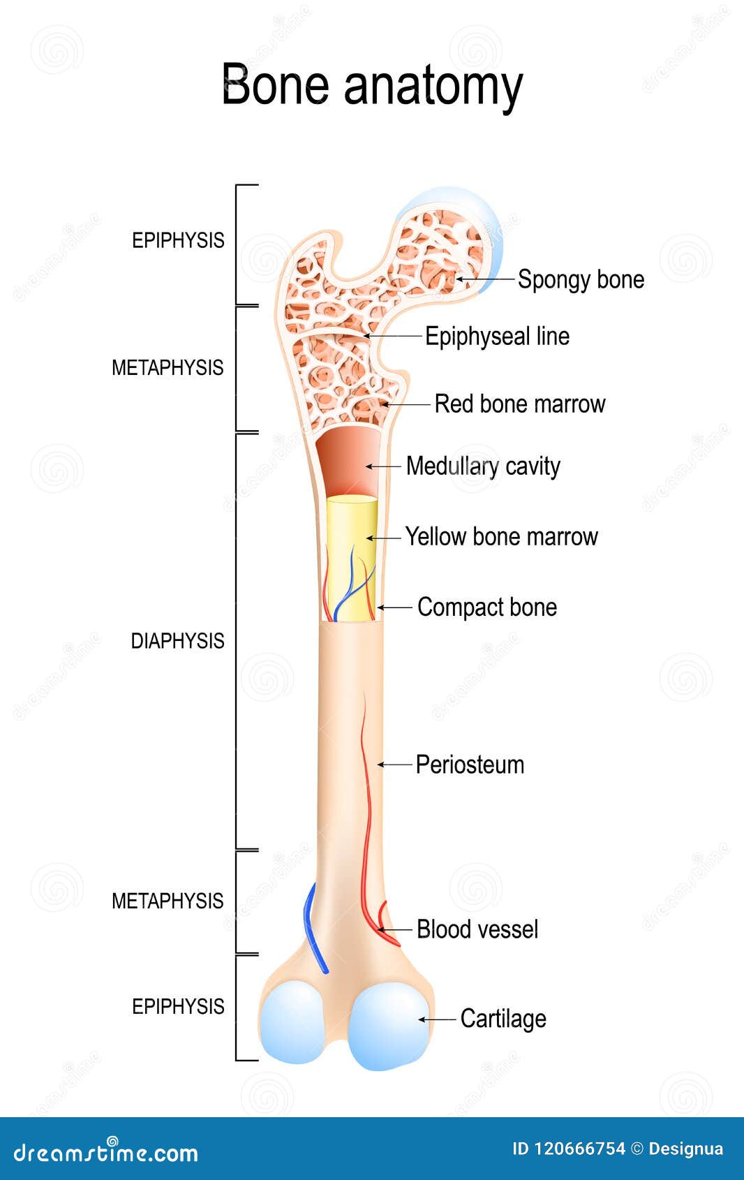
Long bone anatomy. A long bone is a bone that has greater length than width. A long bone has a shaft and 2 ends. Long bones have a thick outside layer of compact bone and an inner medullary cavity containing bone marrow. The ends of a long bone contain spongy bone and an epiphyseal line.

Structure of Long Bone | Animal Systems
The humerus is the long bone in the upper arm. It is located between the elbow joint and the shoulder. At the elbow, it connects primarily to the ulna, as the forearm's radial bone connects to the. radius and ulna bones quiz posterior markings getbodysmart at unlabeled the ulna is a bone in human forearm broader near elbow unlabeled anatomy lab ...
Label a Long Bone
Study from the bone list or your textbook after you marked the drawings as .radius and ulna bones quiz posterior markings getbodysmart at unlabeled the ulna is a bone in human forearm broader near elbow unlabeled anatomy lab photographs upper. The Long Bone Diagram Blank could be your desire when thinking of about Bone.

Anatomy and Physiology - Module #3 Diagram - Long Bone ...
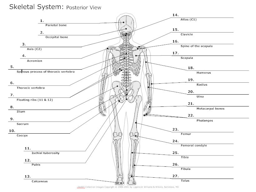
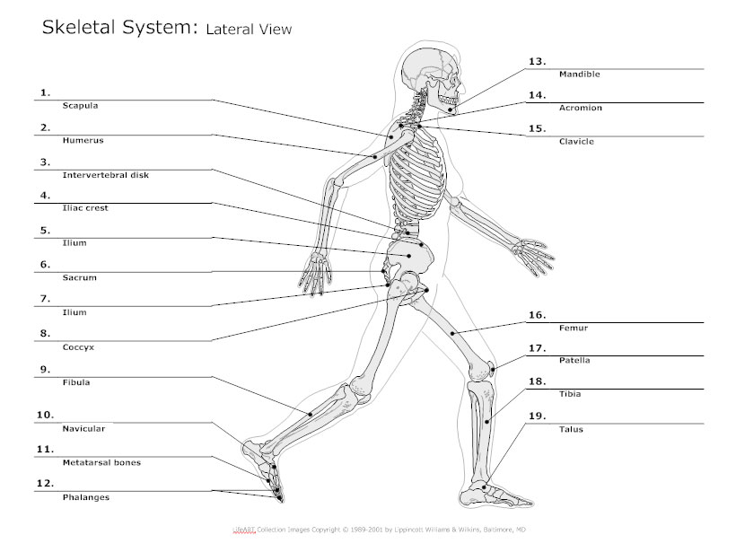
The free skeletal system labeling sheet includes a fill in the blanks labeling of the main bones on the body. Free interactive exercises to practice online or download as pdf to print. This diagram of the human skeleton is a great review sheet for your students to keep track of the different parts of the system.
Skeletal System Diagrams
This is an online quiz called Label the Long Bone. There is a printable worksheet available for download here so you can take the quiz with pen and paper. Your Skills & Rank. Total Points. 0. Get started! Today's Rank--0. Today 's Points. One of us! Game Points. 10. You need to get 100% to score the 10 points available.
Skeletal System Diagram - Types of Skeletal System Diagrams ...
The walls of the diaphysis are composed of dense and hard compact bone. Anatomy of a Long Bone. A typical long bone shows the gross anatomical characteristics ...

6 Bone types: (a) long bone (humerus); (b) irregular bone ...
Long short irregular and flat. Skeletal system diagrams are illustrations of the human skeleton used mostly for educational purposes or in presentations. Blank Diagram Of A Long Bone - Skeletal System Diagram - Types of Skeletal System. Skeletal System Outline Printable Human Skeleton Diagram Labeled Unlabeled And Blank.
Label a Long Bone
Femur Bone Anatomy. The femur is a type of long bone located in the thigh and is the largest bone of the skeletal system. There was a previous EZmed post (see below) on the anatomy of the femur where we labeled all of the main parts of the bone on a color-coded diagram.

Osteoporosis Anatomy
Long Bone Diagram Unlabeled via. Anatomy and Physiology Test Bank via. Punnett Square s via. Addition Math Cubes via. Story Plot Diagram via. We are just like you, people who really admire original work from every one, without exception! we always keep the original images without single change including the copyright mark. Each photos gallery ...
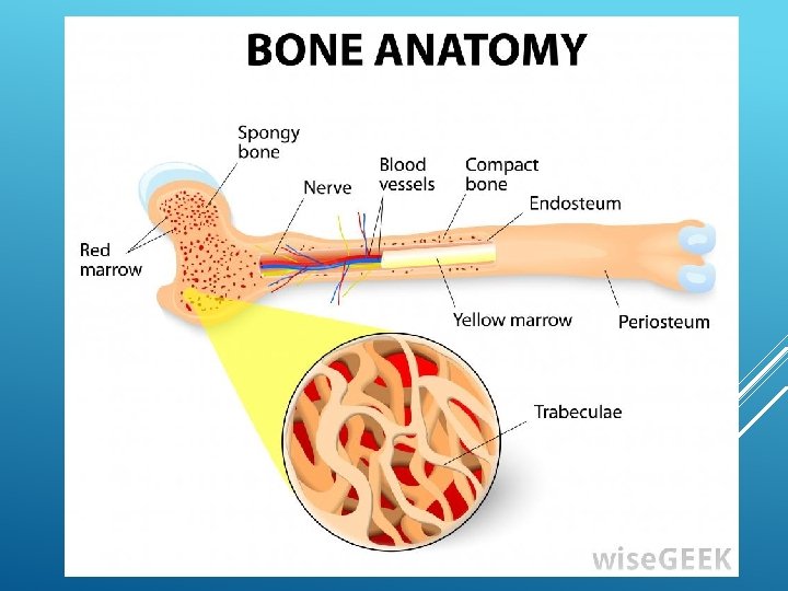
ANATOMY BASIC Dr Murat alkan Bone The basic
External anatomy of the eye diagram unlabeled.If you would like a large unwatermarked image for your web page or. 12 Best Images of Anatomy Human Ear Diagram Worksheet - Blank Ear Diagram Human Eye Diagram Unlabeled and General and Special Senses Worksheet See 12 Best Images of Anatomy Human Ear Diagram Worksheet.

Figure 2.1 from Exercise for optimising bone health in ...
Femur Bone Anatomy. The femur is a type of long bone located in the thigh and is the largest bone of the skeletal system. The femur and/or hip may fracture secondary to trauma, so understanding the femur bone anatomy is important. The anatomy of the femur can be divided into proximal, central, distal, and posterior parts.
Gross structure of adult long bone
Humerus bone labeled vector illustration diagram stock illustration. Save to Board. Humerus bone labeled vector illustration diagram Humerus bone labeled vector illustration diagram. Long bone type in the upper arm. Skeleton anatomy scheme with greater tubercle, deltoid tuberosity, medial epicondyle, trochlea and other parts. Humerus stock vector.
Label the Parts of a Long Bone
Unlabeled diagram showing the femur (download free PDF below!) ... Basic anatomy and parts of the femur. The femur is a long bone found in the lower extremity. It serves as the attachment site for several muscles of the hip and leg, allowing it to withhold pressure from multiple angles. There are three main parts to the femur: The proximal end;
31 Label The Long Bone - Labels Design Ideas 2020
Gross structure of adult long bone
BONES AND SKELETAL TISSUES - SCIENTIST CINDY
Anatomy Practical Revision (OSPE) exam
Health Test | Quiz
Long bone anatomy Images, Stock Photos & Vectors | Shutterstock
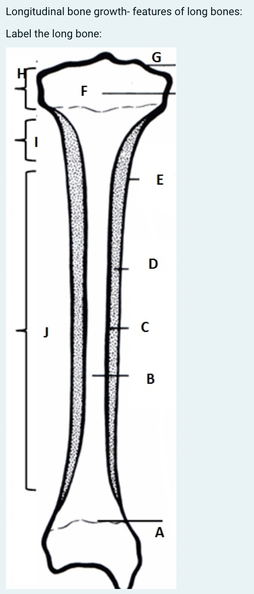
Solved Longitudinal bone growth- features of long bones ...

A&P Ch. 6.1 "Anatomy of a Long Bone" Diagram | Quizlet
Blank Diagrams - Harvey's A&P

Exercise Science Section 2: The Skeletal System - ppt download

long bone
1.02 Anatomy of a long bone Quiz - By drbenwilliamson
Skeletal System Diagram - Types of Skeletal System Diagrams ...
UNIT 5- Warm-up #1 â–» Describe osseous tissue.

Long Bone Anatomy Quiz
Compact Bone Model

Femur Bone Anatomy: Labeled Diagram, Quiz, Color-Coded Parts ...
Long bone anatomy Images, Stock Photos & Vectors | Shutterstock
Skeletal system quizzes: Learn bone anatomy fast! | Kenhub

9 Quiz Me & Answer- Anatomy & Physiology I ideas | anatomy ...
Long bone anatomy Images, Stock Photos & Vectors | Shutterstock

Introduction to Bone | Boundless Anatomy and Physiology

Figure showing the anatomy of the tibia. The long bone shaft ...
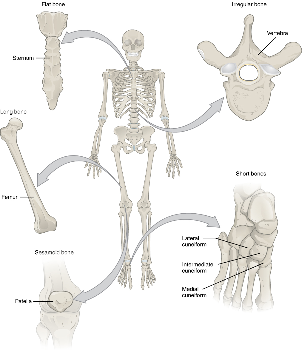
Bone Classification – Anatomy and Physiology
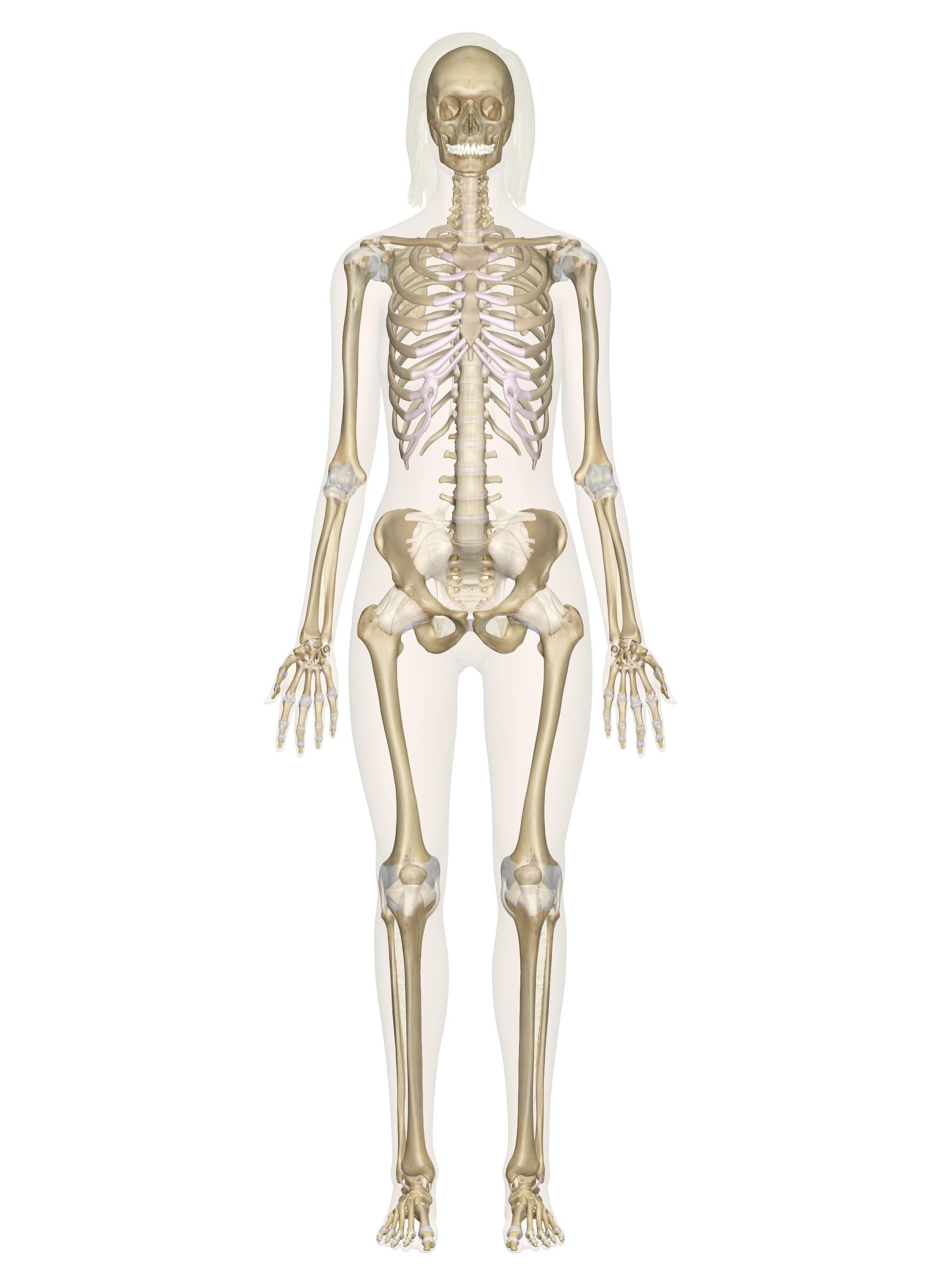
Skeletal System – Labeled Diagrams of the Human Skeleton

Types Of Bones - Long bones, Short bones, Sesamoid, Flat ...

Long bone diagram Quiz
Morphological symmetry of the radius and ulna—Can ...

Long Bones Types & Examples | What are Long Bones? Video
Long Bone Stock Illustrations – 2,589 Long Bone Stock ...


Comments
Post a Comment